Applied Mathematics and Mechanics (English Edition) ›› 2026, Vol. 47 ›› Issue (4): 719-740.doi: https://doi.org/10.1007/s10483-026-3371-9
Previous Articles Next Articles
Haodong WANG1, Ying YU1,†( ), Qingyun WANG1,2
Received:2025-12-12
Revised:2026-02-05
Published:2026-03-31
Contact:
Ying YU, E-mail: yuyingmath@163.comSupported by:2010 MSC Number:
Haodong WANG, Ying YU, Qingyun WANG. Transcranial electrical stimulation enhancing brain network integration: a dynamic modeling study. Applied Mathematics and Mechanics (English Edition), 2026, 47(4): 719-740.

Fig. 1
Whole-brain network model. (a) Each node is implemented as the Jansen-Rit neural mass model, representing a cortical column that comprises three interconnected populations, i.e., pyramidal neurons (the output population), excitatory interneurons, and inhibitory interneurons. The neuronal populations are interconnected via the constants C, C1, C2, C3, and C4. hE and hI represent the impulse response functions. S denotes the sigmoid function. Pyramidal neurons project to other nodes and receive Gaussian-distributed input p(t) as well as inputs from other nodes. (b) The connectivity matrix extracted from the structural data. (c) Nodes are connected through the structural matrix. (d) The dynamic activity of the node output. (e) Based on the generalized hemodynamic model, simulated blood oxygenation level dependent (BOLD)-like recording for the entire brain region is obtained (color online)"
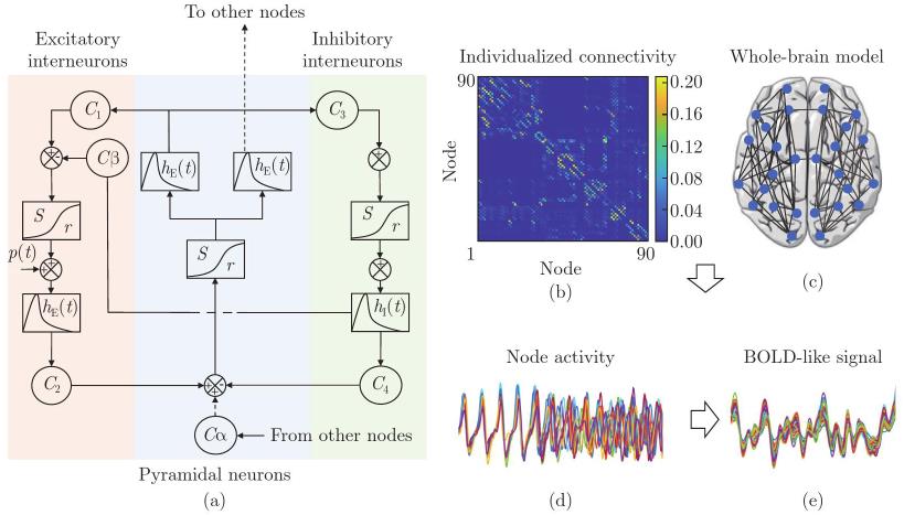
Table 1
Parameter interpretation and values of the Jansen-Rit model"
| Parameter | Definition | Value |
|---|---|---|
| A | Maximum amplitude of the EPSP | 3.25 mV |
| B | Maximum amplitude of the inhibitory post-synaptic potential (IPSP) | 22 mV |
| a | Inverse of the time constant in the feedback excitatory circuit | 100 s-1 |
| b | Inverse of the time constant in the feedback inhibitory circuit | 50 s-1 |
| | Number of synapses on the dendrites of excitatory feedback circuit | |
| | Number of synapses on the dendrites of inhibitory feedback circuit | |
| | Parameters of the sigmoid function | |
Fig. 2
(a)–(c) Characteristics of changes in the average synchronization level R¯, global efficiency EG, and modularity Q across the γβ-parameter space; (d)–(g) node activities, BOLD signals, and sFC matrices for the parameter pairs (γ, β) of (0.4, 0.05), (1.2, 0.15), (2, 0.25), and (3, 0.45), respectively, where the values in the matrix represent the Pearson correlations of BOLD-like signals. As the parameters increase, neurons shift from low-amplitude random oscillations to stable discharge activities, and then to high-amplitude hyperexcited states. To reflect the specific characteristics of discharges at different scales, the node activity signals are displayed within a 5-second period, while the sFC matrix is constructed based on a 600-second time series (color online)"
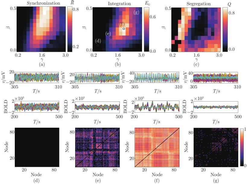
Fig. 3
Electric field distributions and modulation of network dynamics under tES. (a)–(b) Whole-brain electric field intensity distributions obtained via ROAST simulation under different electrode configurations: (a) anode at C3 and (b) anode at C3 and cathode at C4; (c) magnitude of Enormal changes under dual-electrode stimulation; (d)–(e) variations in the average regional synchronization level R¯ with the stimulation intensity under anode and dual-electrode configurations, respectively, where the tES leads to a decrease in the mean synchronization level; (g)–(h) changes in the integration level under stimulation. As the stimulus intensity increases, EG exhibits a non-monotonic trend of first increasing and then decreasing; (f)–(i) EG as a function of both the global coupling parameter and tES intensity (color online)"
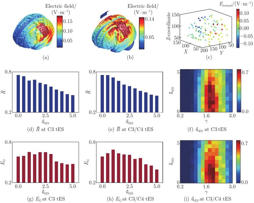
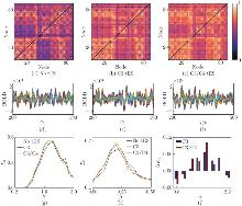
Fig. 4
(a)–(c) Node under three conditions, i.e., no stimulation, anodal tES at C3, and dual-electrode tES at C3/C4, respectively; (d)–(f) sFC and BOLD signals under three conditions, respectively, when γ=1.6 and β=0.2; (g) and (h) EG as a function of the global coupling parameter γ, with β fixed at 0.2, and the inhibitory gain β, with γ fixed at 1.6, where tES enhances integration within specific parameter ranges u^tES=1; (i) change in the maximum EG, quantified as ΔEG=max(EGtES)−max(EGnormal), across β∈ [0, 0.5] under different γ values in the range [0.8, 2.2] (color online)"
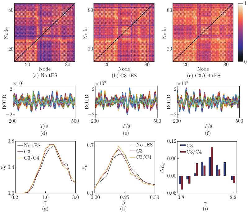
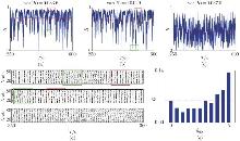
Fig. 5
(a)–(c) Time courses of the phase synchronization level: (a) under no stimulation, (b) stimulation intensity of 1, and (c) stimulation intensity of 5, showing continuous fluctuations between high and low synchronization states, where the corresponding variances are 0.033 6, 0.042 9, and 0.037 0, respectively; (d) raster plot of population firing across brain regions over a 10 s period, where the green box highlights an example of lower synchronization, and the red box indicates a period of higher synchronization; (e) variation in the modularity Q with the stimulation intensity (color online)"
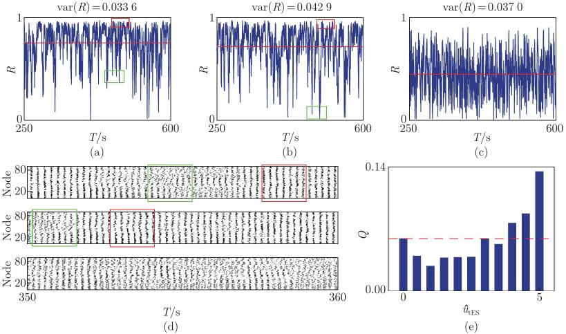
Fig. 6
Whole-brain electric field distributions under different stimulation targets and the corresponding changes in the maximum global efficiency ΔEG: (a) anode at F3 and cathode at F4; (b) anode at FC3 and cathode at FC4; (c) anode at CP5 and cathode at CP6; (d) anode at P3 and cathode at P4, where under different stimulus targets, the maximum value of EG shows a trend of initially increasing and subsequently decreasing with the change in γ, and reaches its peak within the middle range of γ (color online)"
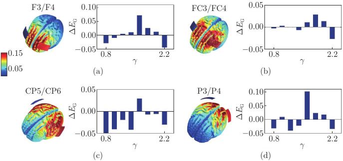
Fig. 7
Effects of focal node lesions on network synchronization and integration. (a) Changes in R¯ and EG (ΔR¯=R¯max,lesion−R¯max,normal, ΔEG=EGmax,lesion−EGmax,normal) after sequentially lesioning each of the 45 left-hemisphere nodes, where error bars indicate the standard deviation of changes; (b) change in the maximum EG following single-node lesions, plotted as a function of the inhibitory gain β (ranging from 0.15 to 0.35), where error bars represent the standard deviation; (c) EG as a function of γ under normal conditions and after lesioning node 7 (selected from the red box in (b), representing the lesion causing the largest decrease in integration), with β fixed at 0.25; (d) EG as a function of γ under normal conditions and after lesioning node 61 (selected from the red box in (b), representing the lesion causing the largest increase in integration), with β fixed at 0.35; (e) sFC matrices at the value of γ yielding the maximum change in EG, where upper panel represents sFC for the normal state versus lesion at node 7 (γ=2), and lower panel represents sFC for the normal state versus lesion at node 61 (γ=2.2) (color online)"
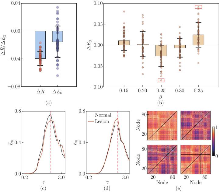
Fig. 8
Recovery of the integration level under tES. (a)–(b) Changes in the integration level under C3/C4 dual-electrode tES at varying stimulation intensities, using node 7 lesion as an example, where red dot line denotes normal level and black dot line denotes lesion level; in (a), the stimulation intensity ranges from 0 to 5; in (b), the range is refined to 0.25–2 to identify the optimal intensity; (c) curves of EG under different conditions, with C3/C4 dual-electrode tES intensity set to 0.25; (d)–(e) changes in the integration level under C3 anodal tES at varying stimulation intensities, ranging from 0 to 5 in (d) and refined to 0.25–2 in (e); (f) curves of EG under different conditions, with C3 anodal tES intensity set to 0.75, where the black, green, and red curves represent the situations of normal, lesion, and tES effects, respectively (color online)"
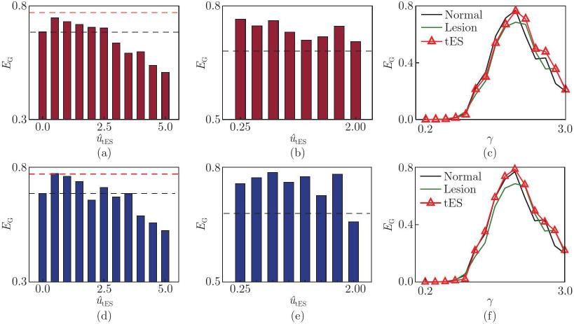
Fig. 9
(a) Changes in EG under different conditions, where the red dots represent the variation in EG after lesioning individual nodes, and the blue and green symbols denote the changes in EG under the C3/C4 dual-electrode tES and C3 anodal tES, respectively; (b) distribution of EG levels across the 45 left-hemisphere nodes (odd numbers from 1 to 89) under individual node lesions is plotted, with the increase in the stimulus intensity u^tES as the vertical axis; (c) impact of node lesions on EG, where the histogram shows the difference in the integration level after lesion relative to the normal state (ΔEG=EGmax,lesion−EGmax,normal); (d)–(e) restorative effect of dual-electrode and single-electrode tES, quantified as the change in EG from the lesioned to stimulated state (ΔEG1=EGmax,tES−EGmax,lesion) (color online)"
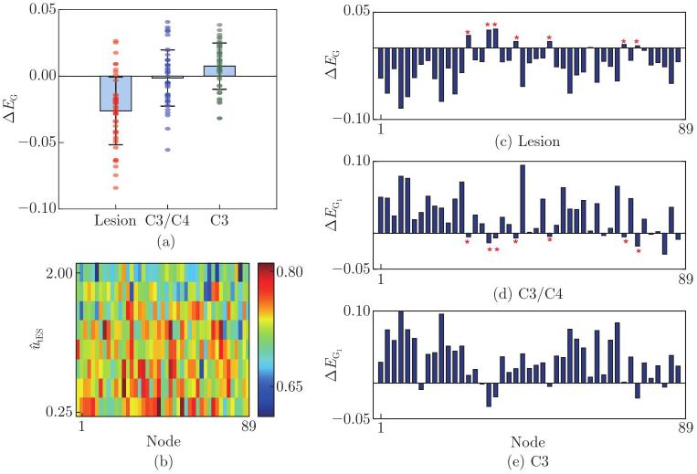
Fig. 10
Effects of diffuse connection loss on network integration and nodal dynamics. (a) Changes in the maximum EG as a function of the global coupling strength γ under 2%, 5%, and 10% connection loss, where for each lesion level, 20 simulations are performed and error bars represent the variance across these runs; (b) representative γ-EG curves under different lesion levels; (c)–(d) nodal firing frequency as a function of γ under different conditions, where the red shaded area indicates regular spiking activity at approximately 4.8 Hz, and the lesion causes this bifurcation point to shift towards a higher value of γ (the red dotted line), indicating that the lesion system requires stronger global coupling to achieve the same dynamic state (color online)"
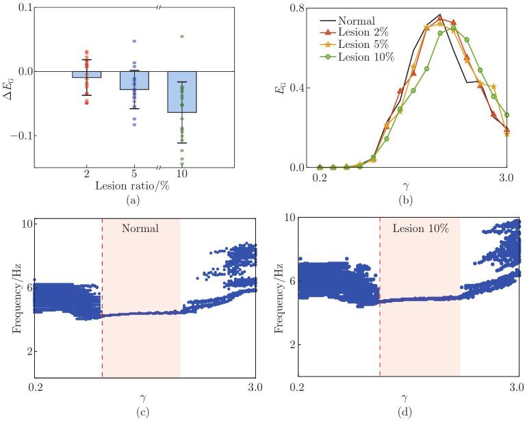
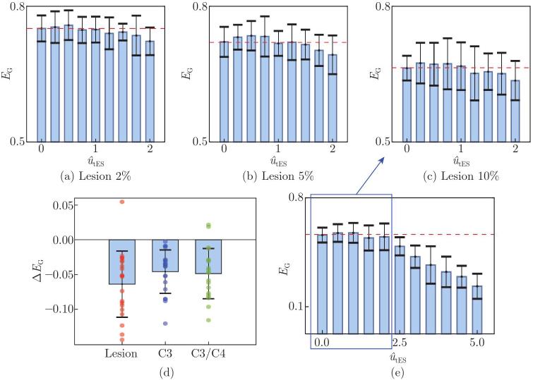
Fig. 11
(a)–(c) Changes in the integration level with the stimulation intensity in the range [0, 2] under the connection loss of (a) 2%, (b) 5%, and (c) 10%, where the red dashed line indicates the integration level without stimulation, and for each lesion level, 20 simulations are performed, in which error bars represent the variance across these runs; (d) difference in the maximum global efficiency EG relative to the normal level under 10% connection loss, comparing conditions without stimulation, with C3/C4 dual-electrode tES, and with C3 single-electrode tES; (e) changes in the integration level with the stimulation intensity in the range [0, 5] under 10% connection loss (color online)"
| [1] | COHEN, J. R. and D’ESPOSITO, M. The segregation and integration of distinct brain networks and their relationship to cognition. The Journal of Neuroscience, 36(48), 12083–12094 (2016) |
| [2] | SHINE, J. M., BREAKSPEAR, M., BELL, P. T., EHGOETZ MARTENS, K. A., SHINE, R., KOYEJO, O., SPORNS, O., and POLDRACK, R. A. Human cognition involves the dynamic integration of neural activity and neuromodulatory systems. Nature Neuroscience, 22(2), 289–296 (2019) |
| [3] | DECO, G., TONONI, G., BOLY, M., and KRINGELBACH, M. L. Rethinking segregation and integration: contributions of whole-brain modelling. Nature Reviews Neuroscience, 16(7), 430–439 (2015) |
| [4] | SHINE, J. M. Neuromodulatory influences on integration and segregation in the brain. Trends in Cognitive Sciences, 23(7), 572–583 (2019) |
| [5] | SHINE, J. M., BISSETT, P. G., BELL, P. T., KOYEJO, O., BALSTERS, J. H., GORGOLEWSKI, K. J., MOODIE, C. A., and POLDRACK, R. A. The dynamics of functional brain networks: integrated network states during cognitive task performance. Neuron, 92(2), 544–554 (2016) |
| [6] | WESTPHAL, A. J., WANG, S. L., and RISSMAN, J. Episodic memory retrieval benefits from a less modular brain network organization. The Journal of Neuroscience, 37(13), 3523–3531 (2017) |
| [7] | REN, S., LI, J. H., TAYA, F., DESOUZA, J., THAKOR, N. V., and BEZERIANOS, A. Dynamic functional segregation and integration in human brain network during complex tasks. IEEE Transactions on Neural Systems and Rehabilitation Engineering, 25(6), 547–556 (2017) |
| [8] | SPORNS, O. Network attributes for segregation and integration in the human brain. Current Opinion in Neurobiology, 23(2), 162–171 (2013) |
| [9] | BREAKSPEAR, M. Dynamic models of large-scale brain activity. Nature Neuroscience, 20(3), 340–352 (2017) |
| [10] | LIU, X. T., YU, Y., HAN, F., ZHOU, J., LIU, Z., LUAN, G. M., and WANG, Q. Y. A data-driven brain network modeling for epileptogenic spread analysis. Biomedical Signal Processing and Control, 105, 107645 (2025) |
| [11] | LIU, X. T., YU, Y., and WANG, Q. Y. Dynamics of epileptic seizure propagation under the regulation of ion mechanisms and synaptic networks. Nonlinear Dynamics, 114, 59 (2026) |
| [12] | CORONEL-OLIVEROS, C., CASTRO, S., COFRÉ, R., and ORIO, P. Structural features of the human connectome that facilitate the switching of brain dynamics via noradrenergic neuromodulation. Frontiers in Computational Neuroscience, 15, 687075 (2021) |
| [13] | YU, Y., FAN, Y. B., HAN, F., LUAN, G. M., and WANG, Q. Y. Transcranial direct current stimulation inhibits epileptic activity propagation in a large-scale brain network model. Science China Technological Sciences, 66(12), 3628–3638 (2023) |
| [14] | YU, Y., WANG, X. M., WANG, Q. S., and WANG, Q. Y. A review of computational modeling and deep brain stimulation: applications to Parkinson’s disease. Applied Mathematics and Mechanics (English Edition), 41(12), 1747–1768 (2020) https://doi.org/10.1007/s10483-020-2689-9 |
| [15] | SUN, Z. K., LIU, Y. Y., YANG, X. L., and XU, W. Control of epileptic activities in a cortex network of multiple coupled neural populations under electromagnetic induction. Applied Mathematics and Mechanics (English Edition), 44(3), 499–514 (2023) https://doi.org/10.1007/s10483-023-2969-9 |
| [16] | SHINE, J. M., ABURN, M. J., BREAKSPEAR, M., and POLDRACK, R. A. The modulation of neural gain facilitates a transition between functional segregation and integration in the brain. eLife, 7, e31130 (2018) |
| [17] | LI, M. K., HAN, Y. N., ABURN, M. J., BREAKSPEAR, M., POLDRACK, R. A., SHINE, J. M., and LIZIER, J. T. Transitions in information processing dynamics at the whole-brain network level are driven by alterations in neural gain. PLoS Computational Biology, 15(6), e1006957 (2019) |
| [18] | CORONEL-OLIVEROS, C., COFRÉ, R., and ORIO, P. Cholinergic neuromodulation of inhibitory interneurons facilitates functional integration in whole-brain models. PLoS Computational Biology, 17(3), e1008737 (2021) |
| [19] | AERTS, H., FIAS, W., CAEYENBERGHS, K., and MARINAZZO, D. Brain networks under attack: robustness properties and the impact of lesions. Brain, 139, 3063–3083 (2016) |
| [20] | LEVIN, M. F., SELLES, R. W., VERHEUL, M. H. G., and MEIJER, O. G. Deficits in the coordination of agonist and antagonist muscles in stroke patients: implications for normal motor control. Brain Research, 853(2), 352–369 (2000) |
| [21] | LI, Y. X., WU, P., LIANG, F. R., and HUANG, W. H. The microstructural status of the corpus callosum is associated with the degree of motor function and neurological deficit in stroke patients. PLoS One, 10(3), e0122615 (2015) |
| [22] | ALSTOTT, J., BREAKSPEAR, M., HAGMANN, P., CAMMOUN, L., and SPORNS, O. Modeling the impact of lesions in the human brain. PLoS Computational Biology, 5(6), e1000408 (2009) |
| [23] | VÁŠA, F., SHANAHAN, M., HELLYER, P. J., SCOTT, G., CABRAL, J., and LEECH, R. Effects of lesions on synchrony and metastability in cortical networks. NeuroImage, 118, 456–467 (2015) |
| [24] | WANG, H. D., JIA, W. L., ZHOU, Y. J., LI, Z. X., YU, Y., and WANG, Q. Y. Unveiling the role of excitation-inhibition homeostasis in stroke recovery from individualized whole-brain dynamical modeling. Communications in Nonlinear Science and Numerical Simulation, 151, 109114 (2025) |
| [25] | HELLYER, P. J., SCOTT, G., SHANAHAN, M., SHARP, D. J., and LEECH, R. Cognitive flexibility through metastable neural dynamics is disrupted by damage to the structural connectome. Journal of Neuroscience, 35(24), 9050–9063 (2015) |
| [26] | LORD, L. D., STEVNER, A. B., DECO, G., and KRINGELBACH, M. L. Understanding principles of integration and segregation using whole-brain computational connectomics: implications for neuropsychiatric disorders. Philosophical Transactions Series A, Mathematical, Physical, and Engineering Sciences, 375(2096), 20160283 (2017) |
| [27] | FLÖEL, A. tDCS-enhanced motor and cognitive function in neurological diseases. NeuroImage, 85, 934–947 (2014) |
| [28] | ORRÙ, G., CONVERSANO, C., HITCHCOTT, P. K., and GEMIGNANI, A. Motor stroke recovery after tDCS: a systematic review. Reviews in the Neurosciences, 31(2), 201–218 (2020) |
| [29] | YUN, G. J., CHUN, M. H., and KIM, B. R. The effects of transcranial direct-current stimulation on cognition in stroke patients. Journal of Stroke, 17(3), 354–358 (2015) |
| [30] | KUNZE, T., HUNOLD, A., HAUEISEN, J., JIRSA, V., and SPIEGLER, A. Transcranial direct current stimulation changes resting state functional connectivity: a large-scale brain network modeling study. NeuroImage, 140, 174–187 (2016) |
| [31] | MERLET, I., BIROT, G., SALVADOR, R., MOLAEE-ARDEKANI, B., MEKONNEN, A., SORIA-FRISH, A., RUFFINI, G., MIRANDA, P. C., and WENDLING, F. From oscillatory transcranial current stimulation to scalp EEG changes: a biophysical and physiological modeling study. PLoS One, 8(2), e57330 (2013) |
| [32] | YU, Y., WANG, H. D., LIU, X. T., and WANG, Q. Y. Closed-loop transcranial electrical stimulation for inhibiting epileptic activity propagation: a whole-brain model study. Nonlinear Dynamics, 112(24), 21369–21387 (2024) |
| [33] | DONG, Y. Q., WEI, J., PENG, S. J., WU, X. R., XU, Y. R., FENG, J. F., ZHANG, J., JIRSA, V., and XIANG, J. Effects of tDCS of the DLPFC on brain networks: a hybrid brain modeling study. PLoS Computational Biology, 21, e1013486 (2025) |
| [34] | GIORDANO, J., BIKSON, M., KAPPENMAN, E. S., CLARK, V. P., COSLETT, H. B., HAMBLIN, M. R., HAMILTON, R., JANKORD, R., KOZUMBO, W. J., MCKINLEY, R. A., NITSCHE, M. A., REILLY, J. P., RICHARDSON, J., WURZMAN, R., and CALABRESE, E. Mechanisms and effects of transcranial direct current stimulation. Dose-Response, 15(1), 1559325816685467 (2017) |
| [35] | IORDAN, A. D., RYAN, S., TYSZKOWSKI, T., PELTIER, S. J., RAHMAN-FILIPIAK, A., and HAMPSTEAD, B. M. High-definition transcranial direct current stimulation enhances network segregation during spatial navigation in mild cognitive impairment. Cerebral Cortex, 32(23), 5230–5241 (2022) |
| [36] | VECCHIO, F., MIRAGLIA, F., RODELLA, C., ALÙ, F., MINIUSSI, C., ROSSINI, P. M., and PELLICCIARI, M. C. tDCS effects on brain network properties during physiological aging. Pflügers Archiv-European Journal of Physiology, 473(5), 785–792 (2021) |
| [37] | JANSEN, B. H. and RIT, V. G. Electroencephalogram and visual evoked potential generation in a mathematical model of coupled cortical columns. Biological Cybernetics, 73(4), 357–366 (1995) |
| [38] | ŠKOCH, A., REHÁK BUČKOVÁ, B., MAREŠ, J., TINTĚRA, J., SANDA, P., JAJCAY, L., HORÁČEK, J., ŠPANIEL, F., and HLINKA, J. Human brain structural connectivity matrices-ready for modelling. Scientific Data, 9(1), 486 (2022) |
| [39] | STEPHAN, K. E., WEISKOPF, N., DRYSDALE, P. M., ROBINSON, P. A., and FRISTON, K. J. Comparing hemodynamic models with DCM. NeuroImage, 38(3), 387–401 (2007) |
| [40] | HUANG, Y., DATTA, A., BIKSON, M., and PARRA, L. C. Realistic volumetric-approach to simulate transcranial electric stimulation: ROAST: a fully automated open-source pipeline. Journal of Neural Engineering, 16, 056006 (2019) |
| [41] | PION-TONACHINI, L., HSU, S. H., MAKEIG, S., JUNG, T. P., and CAUWENBERGHS, G. Real-time EEG source-mapping toolbox (REST): online ICA and source localization. 2015 37th Annual International Conference of the IEEE Engineering in Medicine and Biology Society (EMBC), IEEE, Milan, 4114–4117 (2015) |
| [42] | RAHMAN, A., REATO, D., ARLOTTI, M., GASCA, F., DATTA, A., PARRA, L. C., and BIKSON, M. Cellular effects of acute direct current stimulation: somatic and synaptic terminal effects. The Journal of Physiology, 591(10), 2563–2578 (2013) |
| [43] | LANCASTER, G., IATSENKO, D., PIDDE, A., TICCINELLI, V., and STEFANOVSKA, A. Surrogate data for hypothesis testing of physical systems. Physics Reports, 748, 1–60 (2018) |
| [44] | BENJAMINI, Y. and HOCHBERG, Y. Controlling the false discovery rate: a practical and powerful approach to multiple testing. Journal of the Royal Statistical Society. Series B (Methodological), 57(1), 289–300 (1995) |
| [45] | LATORA, V. and MARCHIORI, M. Efficient behavior of small-world networks. Physical Review Letters, 87(19), 198701 (2001) |
| [46] | RUBINOV, M. and SPORNS, O. Complex network measures of brain connectivity: uses and interpretations. NeuroImage, 52(3), 1059–1069 (2010) |
| [47] | ACEBRÓN, J. A., BONILLA, L. L., PÉREZ VICENTE, C. J., RITORT, F., and SPIGLER, R. The Kuramoto model: a simple paradigm for synchronization phenomena. Reviews of Modern Physics, 77(1), 137–185 (2005) |
| [1] | R. A. JAFARI-TALOOKOLAEI, H. GHANDVAR, E. JUMAEV, S. KHATIR, T. CUONG-LE. Free vibration and transient response of double curved beams connected by intermediate straight beams [J]. Applied Mathematics and Mechanics (English Edition), 2025, 46(1): 37-62. |
| [2] | Chentong GAO, Huiyu SUN, Jianping GU, W. M. HUANG. Dynamic modeling of a three-dimensional braided compositethin plate considering braiding directions [J]. Applied Mathematics and Mechanics (English Edition), 2025, 46(1): 123-138. |
| [3] | Xiaofeng LIU, Qishuai WANG, Haiquan LI, Guoping CAI. Dynamics and control of variable geometry truss manipulator [J]. Applied Mathematics and Mechanics (English Edition), 2017, 38(2): 243-262. |
| [4] | Haiyan SONG, Lifu LIANG. Investigation of power-type variational principles in liquid-filled system [J]. Applied Mathematics and Mechanics (English Edition), 2015, 36(12): 1651-1662. |
| [5] | Wenan JIANG, Guoce ZHANG, Liqun CHEN. Forced response of quadratic nonlinear oscillator: comparison of various approaches [J]. Applied Mathematics and Mechanics (English Edition), 2015, 36(11): 1403-1416. |
| [6] | Fengxiang MEI, Jinchao CUI. Skew-gradient representations of constrained mechanical systems [J]. Applied Mathematics and Mechanics (English Edition), 2015, 36(7): 873-882. |
| [7] | A. M. SIDDIQUI, H. ASHRAF, A. WALAIT, T. HAROON. On study of horizontal thin film flow of Sisko fluid due to surface tension gradient [J]. Applied Mathematics and Mechanics (English Edition), 2015, 36(7): 847-862. |
| [8] | A. FARROKHABADI;A. KOOCHI;A. KAZEMI;M. ABADYAN. Effects of size-dependent elasticity on stability of nanotweezers [J]. Applied Mathematics and Mechanics (English Edition), 2014, 35(12): 1573-1590. |
| [9] | Hui-bin WU;Feng-xiang MEI. Form invariance and conserved quantity for weakly nonholonomic system [J]. Applied Mathematics and Mechanics (English Edition), 2014, 35(10): 1293-1300. |
| [10] | Fang GAO;Xiao-bo ZHANG;Jing-li FU. Application of canonical coordinates for solving single-freedom constraint mechanical systems [J]. Applied Mathematics and Mechanics (English Edition), 2014, 35(8): 1029-1038. |
| [11] | K. REGER;R. A. VAN GORDER. Lane-Emden equations of second kind modelling thermal explosion in infinite cylinder and sphere [J]. Applied Mathematics and Mechanics (English Edition), 2013, 34(12): 1439-1452. |
| [12] | Feng-ming LI;Chun-chuan LIU . Parametric vibration stability and active control of nonlinear beams [J]. Applied Mathematics and Mechanics (English Edition), 2012, 33(11): 1381-1392. |
| [13] | Wei-li HUANG;Jian-le CAI. Conformal invariance for nonholonomic system of Chetaev’s type with variable mass [J]. Applied Mathematics and Mechanics (English Edition), 2012, 33(11): 1393-1402. |
| [14] | Xiu-gen WU;Bai-lin ZHENG;Peng-fei HE;Shu-guang LIU . Equilibrium equations for 3D critical buckling of helical springs [J]. Applied Mathematics and Mechanics (English Edition), 2012, 33(8): 1049-1058. |
| [15] | ZHAO Lu-Hai-Bo;HU Guo-Hui;ZHOU Zhe-Wei. Linear instability of ultra-thin liquid films flowing down cylindrical fibre [J]. Applied Mathematics and Mechanics (English Edition), 2011, 2(32): 133-140. |
| Viewed | ||||||
|
Full text |
|
|||||
|
Abstract |
|
|||||


 Email Alert
 RSS
RSS